Elbow dysplasia is the most common cause of thoracic limb lameness in young, large and giant breed dogs, although small dogs are also affected. Elbow dysplasia is caused by the abnormal association between the humerus, radius and ulna. In many cases, the radius and ulna may not be growing at exactly the same speed, resulting in excessive pressure in certain areas of the joint. Dogs begin to show signs of pain and lameness and to develop osteoarthritis. The abnormal forces on the medial coronoid process of the ulna result in microfractures. A portion of the medial coronoid process separates from the surrounding bone, causing a fragmented medial coronoid process.
At Four Seasons, we use a Vimago 3D imaging scan along with radiographs, to make an accurate diagnosis in a non-invasive way. When a fragmented medial coronoid process is confirmed, elbow arthroscopy (subtotal coronoidectomy) is performed to remove the fragment and surrounding bone. This procedure requires two keyhole incisions at the medial aspect of the elbow, and takes about 30 minutes per elbow. We discharge many patients the same day as surgery and most dogs are able to return to normal activity within six weeks with little or no lameness. The cost for consultation, 3D imaging, radiography, anesthesia and monitoring, bilateral elbow arthroscopy, and hospitalization is approximately $3,000 to $5000.
Although we cannot prevent osteoarthritis in these patients, when diagnosed at a young age (<18 months), before excessive osteoarthritis develops, we have the best chance of helping them long term. In many cases, removing these fragments at a young age and maintaining a lean body condition will slow down the progression of osteoarthritis considerably.
When he first presented to Four Seasons last Fall, Finn was an 11.5 month old MI Border Collie, with five months of intermittent left thoracic limb lameness, especially when trotting up inclines. There was no history of trauma, but there was tenderness at the left medial elbow. Finn received regular chiropractic treatments and laser therapy prior to seeking treatment at Four Seasons.
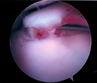
Finn’s radiographs showed early degenerative joint disease was present in the left elbow, suspicious for medial coronoid disease. A Vimago scan of his elbows and shoulders was performed at Four Seasons and revealed bilateral medial coronoid disease. Elbow arthroscopy provided photo documentation of the fragments and the associated cartilage damage, as well as a portal for a subtotal coronoidectomy on each side.
Finn was placed on exercise restriction for six weeks post-operatively and he received twice weekly laser treatments. His recovery was better than expected, according to his owner, who has no regrets for moving forward with surgery. Four months out from surgery, Finn is doing exceptionally well, as the videos below attest. Dancing has replaced agility for him. He has no lameness or residual pain. Finn’s is not an uncommon result.
This video shows Finn approximately 3 months after surgery. He shows no sign of lameness or pain. He moves easily and willingly.
This video shows Finn’s new hobby, dancing, which has replaced the more jarring impact of agility. He’d only had about four dance lessons at the time this video was recorded!